Minimal speakers (MS) represent one-third of the autism spectrum, yet only 2% of autism research participants (Russell et al., 2019). Research conclusions based on people unlike minimal speakers in significant ways may be skewed, leading to profound misunderstanding. We aim to correct underrepresentation of an important group.
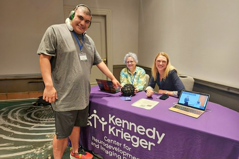
Isaiah points to the HD-DOT cap on display at the 2025 Motormorphosis Conference Kennedy Krieger Institute booth staffed by Karen, on the left, and Morgan.
We agree with Jaswal et al. (2026) that MS “cannot use speech to reliably convey what they want, explain what they know and feel, ask questions, express opinions, or share memories.” MS do not have enough control of their speech muscles to use their voices to maintain autonomy. The neurodevelopmental condition that makes it difficult to perform motor skills, like speaking, is dyspraxia (also known as developmental coordination disorder). This neurodevelopmental disorder begins in childhood, results in acquisition of motor and coordination skills that are below age expectations, significantly interferes with activities of daily living, and is not better explained by another condition like cerebral palsy (American Psychiatric Association, 2013).
Human brains are organized by division of labor. Different parts of our brains are specialized to do different things. We have a motor network that is specialized for planning and executing movement, including speech, and we have a different network responsible for language knowledge and language processing. It is entirely possible for someone to understand everything that is being said to them but be unable to respond because the motor and language networks are anatomically separate. This is an uncontroversial statement in neuroscience, and it means we should never conflate the ability to speak with the ability to understand. And yet, the default assumption is that if an autistic individual is nonspeaking, they have an intellectual disability. One of us is proof that this assumption is wrong. Some MS are intellectually disabled, but we cannot be certain until we have better diagnostic tools. Eleven-year-old Isaiah’s Peabody Picture Vocabulary Test results were “receptive vocabulary currently falls below the first percentile” and “He was able to correctly count pictured objects, could label colours, shapes, and some everyday objects and actions.” The evaluator did not clue in that he had had hundreds of hours of practice pointing to colors and shapes, but not the items he got wrong!
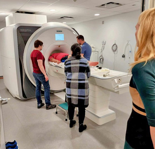
Morgan looks on as Isaiah is positioned for an fMRI scan at Toronto Neuroimaging Facility by MRI Physicist Lars Kasper and MRI Technologist John Milne. Isaiah’s mother, Melody Tien Grewal, provides regulation support.
Neuroimaging
Traditional tests of language comprehension and intellectual ability require someone to speak, point, press a button, or make some other reliable motor response. But for many MS, reliable motor responses are precisely the challenges that make assessment difficult. How can we measure what a person understands if our methods depend on motor abilities that may be impaired?
Neuroimaging offers one way to address this challenge. Rather than relying solely on what a person can physically express, neuroimaging allows researchers to examine how the brain responds while a person listens to language or attempts to perform a task. It provides an important complementary source of information for populations whose abilities may be underestimated by conventional testing. Our work focuses on two methods, functional magnetic resonance imaging (fMRI) and high-density diffuse optical tomography (HD-DOT). These different technologies rely on the same basic principle: when a region of the brain becomes active, it requires additional energy, triggering an increase in local blood flow. This influx of oxygenated blood changes the balance of oxygenated and deoxygenated hemoglobin in that region, creating a blood-oxygen-level-dependent (BOLD) signal. By measuring BOLD signals while a person performs an active or passive task, researchers infer which brain regions are involved. Both methods allow researchers to study the neural systems that support language, cognition, and movement without requiring extensive motor responses.
Our team from the Kennedy Krieger Institute, the University of Toronto, MGH Institute of Health Professions, Washington University Medicine, and Stony Brook University recently co-designed two neuroimaging studies, working closely from the outset with minimally speaking community members to develop tasks that would both be feasible for people with dyspraxia and yield rigorous scientific data. Our minimally speaking colleagues helped shape many aspects of the study design, such as limiting imaging sessions to one hour, including time to become comfortable with the equipment and practice the experimental tasks. Legal guardians or participants themselves provided informed written consent. Data collection began only after each participant indicated they were comfortable proceeding.
Functional Magnetic Resonance Imaging (fMRI) – fMRI is one of the most powerful tools for studying the human brain because it can measure activity throughout the entire brain, including deep structures that cannot be accessed using HD-DOT. Whole-brain coverage is particularly important when studying language and motor function because these abilities depend on networks of regions, some deep inside the brain, working together.
But fMRI presents substantial challenges for many MS. Participants must lie still inside a narrow scanner while loud sounds are produced by the MRI system. Even small head movements can degrade data quality. To make participation possible, we worked closely with MS and their families to develop accommodations that reduced sensory and motor demands. We closed our facility so that only the minimally speaking participants, their family members, and the research team were present. Family members accompanied participants into the scanner room to hold their hand or provide regulation support during the scan. Participants had repeated opportunities to enter and exit the scanner, practice the tasks, and decide whether they wished to continue. We also used state-of-the-art motion-correction technology to reduce the impact of movement during scanning.
Using these approaches, we have successfully localized language networks in two minimally speaking autistic adults, providing some of the first neural evidence of language processing in this population. These preliminary findings demonstrate that meaningful fMRI research with MS is possible and underscore the importance of developing methods that allow MS to be included in research on complex cognition. Data collection is ongoing.
High-Density Diffuse Optical Tomography (HD-DOT) – HD-DOT is a recent imaging technique that promises to be more accessible for MS. In HD-DOT, a participant wears a cap that contains infrared light sources and detectors. Infrared light shines through the scalp and brain in the same way that the light in a pulse oximeter worn on the finger does. The cap’s detectors then detect the light after it has passed through these tissues. HD-DOT is easier to tolerate than MRI because people have some freedom to move. The cap is tight-fitting, but unlike EEG caps, which use gel or saline solution to create a connection between electrodes and a person’s scalp, it is dry, making it a more feasible option for people with sensory sensitivities. But the trade-off is that near-infrared light can penetrate only the outer few centimeters of the brain. We can measure activity in the cerebral cortex but not in deeper brain structures.
Our HD-DOT participants performed three tasks, taking breaks as needed: repetitive and sequential finger tapping, repeating single and multiple syllables, and listening to understandable and acoustically degraded speech. Recognizing that the motor tasks and the feel of the cap are very challenging for many MS, we provided participants with visual and written cues and checked in after each task to see how they were doing and confirm whether they wanted to continue or not.
Data from 17 nonspeaking participants passed our data quality checks, showing good contact between the cap and scalp, and that the amount of movement was acceptably low. One person discontinued because the cap was too uncomfortable, and not everyone completed all the tasks before tiring out. Compared to a group of 28 non-autistic participants, the brain activation patterns of our MS participants showed similar patterns, but much more variation, suggesting that, on average, they used more of their brains to perform the tasks than the non-autistic group. This is consistent with the idea that these tasks are feasible but harder for MS. “Crucially, we also found that when listening to sentences, many minimally speaking individuals show patterns of brain activation consistent with understanding language content” (CNIR 2025 Research Update).
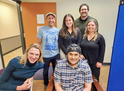
Isaiah takes a break from HD-DOT data collection surrounded by (left to right) Morgan; Dr. Stewart Mostofsky, Director, Center for Neurodevelopmental and Imaging Research, Kennedy Krieger Institute (KKI); Alice Sperry, KKI; Adam Eggebrecht, Associate Professor of Radiology, Washington University Medicine Mallinckrodt Institute of Radiology; Deana Crocetti, KKI
Motor for Research
Here’s Isaiah’s advice for families, nonspeakers, and researchers:
- Having dyspraxia means every intentional action must be practiced. The more an autistic dyspraxic person practices motor control, the more they will be able to control their body to participate in research.
- fMRI work feels like going into a small, short, round container. The best way to practice for the experience is to crawl into similar-feeling spaces in playgrounds, sensory gyms, and one’s bedroom (think of lying down on the floor of a closet with your feet sticking out or under a sheet). If available, practice in a mock scanner. If needed, practice over many sessions to build tolerance.
- HD-DOT work feels like wearing a heavy, hot cap of sci-fi tiles while tethered to a heated cable bundle connected to a computer. The best way to practice for the experience is to wear different types of tight hats for longer and longer periods of time; think toques, baseball caps, and caps worn by swimmers and marathon runners.
- Research staff who want to work with MS must also practice. Run drills for setting up participants and experiments. Nonspeakers have limited reserves of regulation energy, so every minute lost to fiddling with equipment is a minute lost of data collection.
Looking Forward
Our two feasibility studies are far from definitive, but they are an important first step. We show that MS are very interested in research collaboration. We show that HD-DOT may be more tolerable for MS than fMRI. And our data supports previous work that minimally speaking autistic people’s brains are wired differently from the brains of non-autistic people — they may be using different areas of their brains to accomplish the same tasks. Future work will tell us more about the range of variation in minimally speaking people’s brain activation and help us understand why speaking or making other intentional movements are so difficult for some MS.
However, imaging studies can only supplement direct tests of language comprehension. Traditional language tests are not well-suited for people with dyspraxia and typically underestimate their abilities. We need to think carefully and work closely with MS and other dyspraxic people to create tasks that make it possible for them to show all that they know.
Isaiah Tien Grewal is a Trainee in the Stony Brook University School of Social Welfare LEND (Leadership Education in Neurodevelopmental Disabilities) Fellowship Program. Morgan D. Barense, PhD, is Professor and Glassman Chair in Neuropsychology in the Department of Psychology at the University of Toronto and Senior Scientist at the Rotman Research Institute, Baycrest Hospital. Karen Chenausky, PhD, CCC-SLP, is Associate Professor at the MGH Institute of Health Professions. Correspondence may be directed to Isaiah Grewal at [email protected] and Karen Chenausky at [email protected].
References
American Psychiatric Association. (2013). Diagnostic and statistical manual of mental disorders (5th ed.). American Psychiatric Publishing.
Center for Neurodevelopmental and Imaging Research (2025). Research Update https://www.kennedykrieger.org/research/centers-labs-cores/center-for-neurodevelopmental-and-imaging-research/research-briefing.
Jaswal, V. et al. (2026) Why we need to study assisted methods to teach typing to nonspeaking autistic people. Autism Research 19e70176 https://doi.org/10.1002/aur.70176.
Russell, G. et al. (2019) Selection bias on intellectual ability in autism research: A cross-sectional review and meta-analysis. Molecular Autism 10:9 https://doi.org/10.1186/s13229-019-0260-x.